CoLabs supports UCSF and external investigators to use and develop various optical microscopy techniques and image analysis methods.
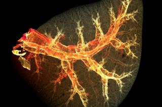
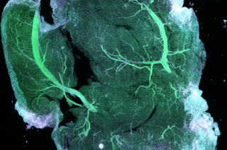
Courtesy of the Looney Lab
CoLabs trains scientists to use our large collection of microscopes and assist with imaging experiments from design to analysis. Visit the Parnassus Advanced Light Microscopy (PALM) CoLab (formerly BIDC) website for more details. Recharge rates are available on iLab.
To get started, please contact PALM. We offer support in fluorophore selection, sample preparation, instrument configuration, and data visualization and analysis. From there, you may work with additional CoLabs Teams depending on your project. Along with optical microscopy, you can take advantage of the complementary platforms, expertise, and collaborative culture at CoLabs. Our labs are located near each other to streamline communication and workflows.
Working within the CoLabs Community
If you are interested in spatial transcriptomics or spatial proteomics, please visit our Spatial Omics platform section. We work with Disease to Biology, Genomics, and Flow Cytometry CoLabs to prepare samples, run slides, and link images with other types of data. To combine image data with other data, contact Data Science CoLab to discuss options for data integration and analysis. For large-scope projects, you can establish a multidisciplinary collaboration (“CoProject”) between you and multiple CoLabs Teams. CoProjects is also a conduit for CoLabs to develop customized methodologies.
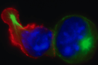
CoLabs maintains and offers a variety of optical microscopes with diverse capabilities:
- inverted and upright
- environmental chamber for live cells and animals
- slide, dish, plate, or custom-made sample mounts
- multiple laser lines, objective magnifications and immersions, tunable filters, and detectors
Many configurations are set up for specialized applications, including:
- laser scanning confocals capable of multiplex imaging
- super resolution spinning disk for live cells and optogenetics
- widefield for thin samples such as cell cultures
- multiphoton for imaging deep into thick tissue
- light sheet for large volume samples
In addition, we have instruments for sample preparation such as vibratomes for sectioning and a micropipette puller for injections.
Scientists may use these instruments independently after appropriate training or work with PALM on their imaging projects. We are happy to help you to choose the best instrument to fit your imaging needs and optimize the microscope configurations for analysis requirements.
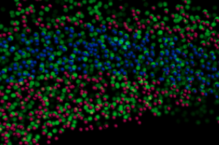
Workstations are available for reservation with installed software including Imaris and Huygens. These are equipped with large, multi-monitors and high-end CPUs and graphics card capable of fast processing and image rendering. Our workstations are helpful for image processing, visualization, and analysis such as deconvolution, colocalization, segmentation, and more.
We also offer image analysis consultations, software training, and collaborations to customize scripts, especially for multi-dimensional datasets.
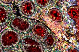
Courtesy of the Molofsky Lab
All users receive individual training sessions for each microscope, analysis station, or piece of support equipment they wish to use. These sessions are tailored to each user’s experience, aptitude, and overall comfort with the instrument. For our more advanced microscope systems there can be 2-4 training sessions including supervised experiments before unaccompanied access is granted.
We also offer a microscopy minicourse and workshops throughout the year. Topics range from the fundamentals of optical microscopy to the latest technological advancements. Please check our Events page for upcoming seminars, webinars, demos, and more.
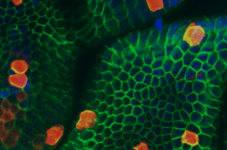
Courtesy of the Kattah Lab
PALM collaborates with researchers to write custom analysis for their image datasets in a variety of platforms including ImageJ, Python, and MATLAB. These codes range from post-processing corrections to tracking and are available here.
1. Where are your microscopes located?
Many of our instruments are located at UCSF Parnassus Heights on the 11th floor of the Medical Sciences Building. We also have microscopes on MSB 6th and 8th floors and in the Broad Stem Cell Center.
2. What if I don’t see an instrument that can do what I need for my research? Can CoLabs manage microscopes purchased by UCSF labs or programs?
The PALM CoLab is enthusiastic about partnering with UCSF investigators and programs to obtain and manage additional microscopes that augment our capacity and bring novel capabilities to UCSF research community. We work with various companies to demo new microscopes and software and often open this trial period to the UCSF community. Please contact us to discuss options.
3. Can someone help me to customize a platform, image analysis program, etc?
We welcome collaborations that build new and/or expand on current technologies. Some of our previous projects can be found here.
4. Who do I contact for more information on training, questions about instrument capabilities, help with image analysis, etc?
palm@ucsf.edu
Medical Sciences Building S1109
513 Parnassus Ave (map)Additional information can be found at the PALM website.


